Citation Information
Antani, JoLS-Pub J Life Sci. Vol.2, No.5, May 2025:1-6 (https://doi.org/pqns)
Introduction
We don’t like bacterial infections. Antibiotics are amazing at treating bacterial infections - when they work. Bacteria rapidly become resistant to antibiotics because they grow and mutate really fast. Over the last century, harmful bacteria have become resistant to our antibiotics. It is very difficult to make new antibiotics. Therefore, antibiotic resistance is becoming a threat to public health. Fear not though, we scientists are coming up with solutions to this problem. One of these solutions is using viruses that attack bacteria- after all, the enemy of our enemy is our friend!
Viruses can infect many different things. You know how the flu virus and COVID-19 infect humans? Well, some viruses actually infect and kill bacteria! Want to see this in action? Check out this YouTube short!. These viruses are called bacteriophages (“bacteria eaters”) or simply “phages”. These viruses are like specialized hunters in nature - just as a bird of prey might only hunt mice or fish, these viruses only hunt specific bacteria. They do not kill human cells. This means we can use them to treat patients with bacterial infections. Scientists call this “phage therapy”, and it’s showing promising results1,2. Bacteria can become
resistant to phages, just like they do with antibiotics. But here's the good news: phages are everywhere in nature! Just like how you can find different types of insects in any backyard, park, or forest, we can find different types of phages in ponds, sewage, the ocean, or soil. And even better: when bacteria try to fight against phages, they often become weaker, which makes them easier to kill with antibiotics3.
Before we use anything to treat patients, we need to understand exactly how it works. This is especially true for phage therapy. That is why we are studying phages in the laboratory. As part of this research, we recently developed a technique to measure how good phages are at grabbing onto bacteria4. Think of it like determining how good a hunter is at catching their prey. When a phage, our hunter, attacks a bacterium, it follows a specific game plan. First, it needs to find and stick to special molecules on the bacterial surface - kind of like finding a door handle to grab onto. Then, it injects its genetic instructions (like a computer program) into the bacterial cell. Inside the cell, the phage makes many copies of itself. Finally, the bacterial cell bursts open, releasing all the new phages to hunt for more bacteria (Fig 1A).
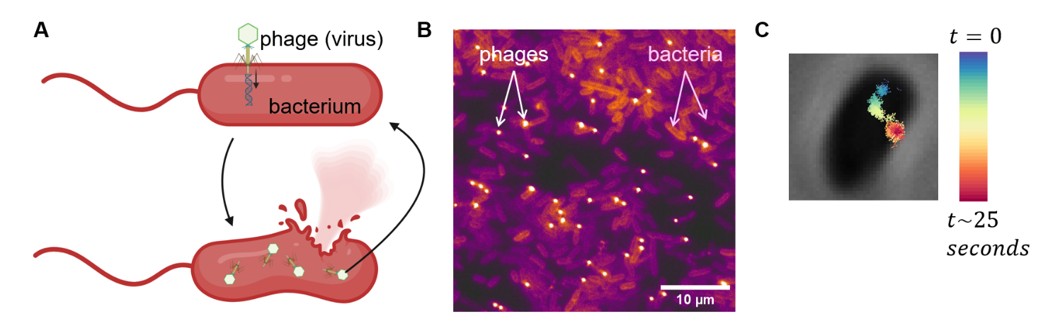
Fig 1. Bacteriophages (phages) are viruses that infect bacteria. (A) The phage life cycle: Like ships finding their home port, phages must first attach to specific spots on bacterial cells. Once docked, they inject their genetic material into the bacteria. Inside the cell, they create many copies of themselves until the bacterial cell bursts, releasing new phages that search for fresh bacterial cells to infect. (B) Microscope image showing phages (bright spots) interacting with bacterial cells (appearing as dimmer rod-shaped objects). (C) By taking multiple images like the one in (B), we can record a movie and track phage movements over time. This example shows a single phage's path as it enters our microscope's view (blue, t = 0 seconds) and moves around for about 25 seconds (color changes from blue to red along its path).
The Mystery: How do viral hunters find their target?
Scientists have been using the same method to study phages for over 100 years. Imagine you're trying to figure out how good people are at finding seats in a crowded cafeteria. The old way would be like counting how many people are still standing every few minutes - it tells you how many found seats, but not how each person actually searched for their spot. Similarly, the old method just mixes phages and bacteria together and counts how many phages haven't found their bacterial targets yet. This is super expensive - like having to prepare a fresh cafeteria full of food every single time you want to do the counting! It also takes forever and needs multiple people working non-stop - imagine having to prepare fresh food, set up the cafeteria, AND count standing people every few minutes during lunch hour! Plus, after all this expensive and time-consuming work, we only learn the average success rate. We still don't know what each individual phage does while searching for its target bacterium.
Microscopy to the rescue!
We wanted to develop a way to watch individual phages as they attach to bacteria. We decided to look at phages under a microscope. Phages have a distinctive shape - a geometric head and legs like a tiny spider (Fig 1A). You can see amazing pictures of these shapes if you google "bacteriophage electron microscopy"! But there's a catch - to take those detailed pictures, scientists must freeze the phages completely still. Since we wanted to watch phages moving around on bacterial surfaces, we needed to use a different kind of microscope that works with light (instead of electrons). In other words, electron microscopes give us really pretty but still photographs, whereas we wanted a live video feed, so we used light (optical) microscopes.
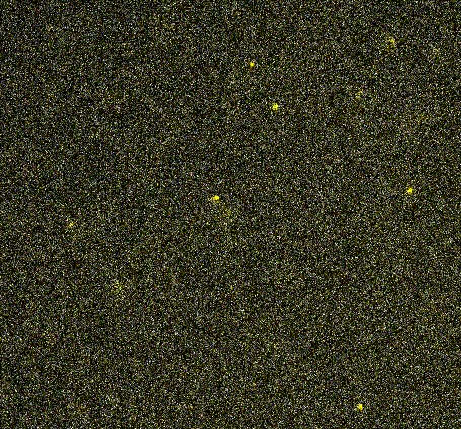
We used a fluorescent dye to color the phages – this is the “science version” of how you would dye your hair or tie-dye a shirt. Just like highlighting important text makes it stand out on a page, fluorescent dyes make our phages light up and shine under the microscope. We could tell the glowing phages apart from the bacteria, which we had stuck onto a glass slide (Fig 1B). We recorded videos of the phages as they moved around (or got stuck) on the bacterial cells – they appeared like glowing dots moving around (see Movie 1). Then we used computer programs to analyze these videos and figure out exactly where the phages went and how long they stayed there. An example is shown in Fig 1C where we trace a phage’s path over time, drawn right on top of a picture of the bacterial cell.
Movie 1. Example time-lapse video of fluorescent phages.
Connecting the Dots: What our data tells us
After collecting all this data, we focused on one key question: how long does each phage spend interacting with bacterial cells? Take a look at Fig 2, which explains what we actually observe in the microscope. The microscope can only show us a thin slice of the sample, like looking through a window into a room. This viewing area is shown as the yellow-shaded area, and we can only see colored phages when they're in this area. Different phages behave in different ways. Some phages (like A and B) only show up briefly or for a medium amount of time. Others (like phage C) stick to cells for a long time. Some phages (like D) never even enter our view through the microscope (shown as the green-shaded area in the bottom rectangle), so we don’t see these phages. You can see examples of these in Movie 1.
We tracked thousands of such phages, including ones moving in and out of our view. Here's the key insight: if a phage sticks strongly to bacteria, we'll see more cases like phage C - long periods where we can see it. By calculating the average time all these thousands of phages spend in view, we can figure out how strongly they typically interact with cells. The stronger they bind, the longer the average time.
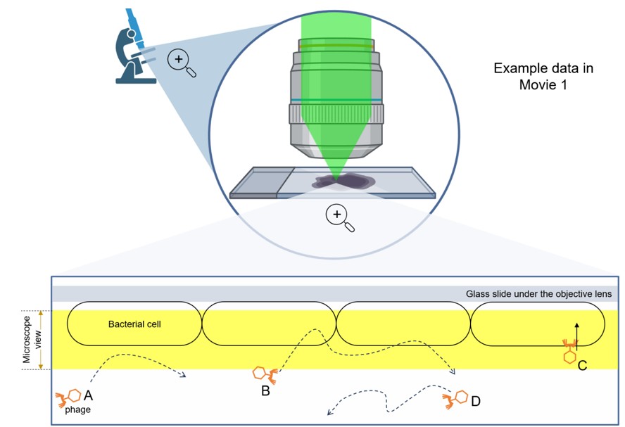
Fig 2. How our microscopy technique works: Watching phages interact with bacteria. The microscope setup is like a spotlight system for watching ships in a harbor. A light beam (shown in green) shines down from above through an objective lens, which focuses the light onto our sample. When this light hits our fluorescent phages, they glow and send light back through the same path to a camera that records their movements. The microscope can only see a thin slice of the sample (shown as a yellow-shaded rectangle). A phage is only visible when it enters this thin field of view. We can imagine four different scenarios:
Phage A: A quick pass-by. Like a ship that briefly crosses through the harbor without stopping, this phage enters and exits our viewing window quickly without interacting with any bacteria. It appears as just a brief flash to our camera.
Phage B: The window shopper. This phage enters our view and explores several bacterial cells (like a ship checking multiple docking stations) before leaving. It stays visible longer than Phage A.
Phage C: The successful dock. This phage finds a bacterial cell and sticks to it (like a ship successfully docking), beginning the infection process. It remains visible throughout our entire observation period.
Phage D: The outsider. This phage never enters our viewing window - like a ship that stays in deeper waters without entering the harbor. Our camera never sees it.
Phages bind to specific molecules (proteins or sugars) that stick out from the bacterial surface, like a ship docking into its assigned spot in the harbor. Just as different types of ships need different kinds of docks - a cruise ship can't dock at a small fishing boat slip - different types of phages need their own specific receptors. These molecules are called "phage receptors". Using genetic engineering, we can remove the genes that make these receptors - kind of like removing the docks from the harbor. Without these receptors, the phages have nothing to grab onto, so they can't bind or infect the cells.
To test if our microscopy idea works, we did a simple comparison. We looked at phages interacting with two types of bacterial cells: normal cells with their receptors intact, and engineered cells with their receptor genes removed. Our prediction was simple: phages should hang around longer near cells that have their receptors (where they can bind) compared to cells without receptors (where they cannot bind). When we performed the experiment, that's exactly what we saw (Fig 3)!

Fig 3. Measuring phage 'stickiness': How long do they stay in our view? We calculate how long phages are visible under the microscope. When phages find bacteria with matching docking sites (receptors), they stick around - like ships finding their correct berths in a well-equipped harbor. We observe these phages for long periods because they attach firmly to the bacterial cells. However, when phages meet bacteria lacking the proper receptors (a harbor with no suitable docks), they quickly drift away. It's like ships approaching a harbor but finding no place to dock - they briefly pass through our view and then disappear, resulting in much shorter average observation times.
In the rest of our scientific paper4, we tested how well this method worked with different combinations of phages and bacteria. We tried many different species of each, including some notorious bacteria known for becoming resistant to antibiotics. Our approach successfully worked for most phages and bacteria that we tested. However, we did find some limitations. Some phages were just too tiny to see with our regular microscope! To see these extra-small phages, we would need more powerful microscopes that use focused laser beams instead of the regular bright light that we had used in our successful experiments.
What's Next? From Lab Bench to Hospital Bedside!
Our findings advance science in two important ways. First, we've developed a method that lets us watch how individual viruses dock with bacteria in real time. This is like having a harbor master's camera system that can track exactly how each ship finds and connects to its dock, helping us understand the details of virus-bacteria interactions.
More importantly, this method has practical applications in medicine. Imagine a patient with a bacterial infection. We might have ten different phages that could potentially fight this infection. Our microscopy technique can quickly show us which phage binds most strongly to the dangerous bacteria, helping us choose the best one for treatment. This is much faster than traditional methods, allowing for quicker decisions in treating infections.
Looking ahead, we're working to make this technique more user-friendly. Our goal is to ensure that even people without extensive microscopy or coding experience can use these tools - like creating an automated harbor monitoring system that anyone can operate.
Notes
- Parts of figures in this article were created with BioRender.com.
- Language editing and accessibility review was performed using Claude 3.5 Sonnet (Anthropic).
References
1. Pirnay, J.-P. et al. Personalized bacteriophage therapy outcomes for 100 consecutive cases: a multicentre, multinational, retrospective observational study. Nat Microbiol 9, 1434–1453 (2024).
2. Zimmer, C. A virus, fished out of a lake, may have saved a man’s life — and advanced science. STAT https://www.statnews.com/2016/12/07/virus-bacteria-phage-therapy/ (2016).
3. Oromí-Bosch, A., Antani, J. D. & Turner, P. E. Developing Phage Therapy That Overcomes the Evolution of Bacterial Resistance. Annual Review of Virology 10, null (2023).
4. Antani, J. D., Ward, T., Emonet, T. & Turner, P. E. Microscopic phage adsorption assay: High-throughput quantification of virus particle attachment to host bacterial cells. Proceedings of the National Academy of Sciences 121, e2410905121 (2024).
| Author | ||
 |
||
| Jyot Antani | ||
| Postdoc Reviewer | Grad Reviewer | Undergrad Reviewer |
 |
 |
 |
| Roya Bagheri | Parisima Ghaffarian | Neha Ramasastry |