Tau oligomers have recently emerged as the principal toxic species in Alzheimer’s disease (AD) and tauopathies. Tau oligomers are spontaneously self-assembled soluble tau proteins that are formed prior to fibrils, and they have been shown to play a central role in neuronal cell death and in the induction of neurodegeneration in animal models. As the therapeutic paradigm shifts to targeting toxic tau oligomers, this suggests the focus to study tau oligomerization in species that are less susceptible to fibrillization. While truncated and mutation containing tau as well as the isolated repeat domains are particularly prone to fibrillization, the wild-type (WT) tau proteins have been shown to be resistant to fibril formation in the absence of aggregation inducers. In this review, we will summarize and discuss the toxicity of WT tau both in vitro and in vivo, as well as its involvement in tau oligomerization and cell-to-cell propagation of pathology. Understanding the role of WT tau will enable more effective biomarker development and therapeutic discovery for treatment of AD and tauopathies.
Keywords: Tau accumulation and toxicity, Tau oligomers and aggregates, Conformational ensembles, Cell-to-cell propagation, Neurodegenerative diseases
Introduction
Alzheimer’s disease (AD) is the 6th leading cause of death in the United States, and there are more than 25 million people suffering from AD worldwide. While several hypotheses have been proposed to elucidate the disease mechanisms, the exact cause of AD pathology is still unknown and remains to be investigated. Wild-type (WT) MAPT gene encoding for microtubule-associated protein tau is carried by most patients with tauopathy, and aberrant accumulation of WT human tau is a hallmark of sporadic AD 1, 2. Tau is an intrinsically disordered protein and a microtubule binding protein that plays an important role in the regulation of microtubule stability and axonal transport 3. Under pathological conditions, tau is hyperphosphorylated and detached from microtubules, and it undergoes misfolding and conformational changes. Cytosolic accumulation of tau triggers the fibrillogenesis pathway, with monomers spontaneously forming into oligomers in the initial stage followed by subsequent fibrillization into intracellular paired helical filaments (PHFs) and neurofibrillary tangles (NFTs) (Fig. 1A)4.
While the presence of the large insoluble NFTs in the brain have been previously suggested to be the main cause of cognitive impairment, recent studies suggest that the spontaneously formed soluble tau oligomers prior to fibrillization contain the principal toxic tau species 5, 6, 7, 8, 9, 10, 11, 12. These toxic tau oligomers induce neuronal cell death as well as neurodegeneration and AD pathology in animal models including mice, Drosophila and Caenorhabditis elegans (C. elegans). While tau proteins may contain mutations or undergo other pathogenic processes such as truncation, and specific regions of tau (e.g. repeat domains) have been identified to be more aggregation-prone 13, the focus of this review will be on WT tau proteins which are less susceptible to fibrillization and mainly involved in the early oligomerization process. We will specifically discuss the critical roles of WT tau in AD pathogenesis, including tau-induced toxicity, formation of tau oligomers as well as their involvement in cell-to-cell propagation of disease pathology (Fig. 1B).
Results
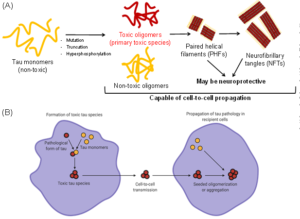
Fig. 1. Tau oligomerization/aggregation cascade and cell-to-cell propagation.
(A) The intrinsically disordered tau monomer is capable of forming tau oligomers spontaneously and producing both toxic and non-toxic soluble tau species. Subsequently, tau oligomers are able to form large insoluble aggregates with -sheet structures, including paired helical filaments (PHFs) and neurofibrillary tangles (NFTs). PHFs and NFTs may be neuroprotective by sequestering toxic oligomers. (B) The toxic tau species (oligomers or aggregates) can be secreted by cells and transmitted to nearby recipient cells. In recipient cells, the uptake of toxic species can further induce seeded oligomerization or aggregation, resulting in cell-to-cell propagation of disease pathology. Schematics were created with BioRender.com.
Tau toxicity
The gain of the toxic function of tau has been primarily attributed to the formation of toxic tau oligomers 10, . The toxic tau oligomers have been postulated to cause cellular dysfunctions such as mitochondrial impairments and apoptosis induced by activated caspase, both of which impede synaptic energy production and result in cell death 15, 16. These oligomers can be either on- or off-pathway, where on-pathway corresponds to their ability to subsequently form fibrils, and off-pathway does not fibrillize. In addition, the tau oligomers are capable of inducing tau misfolding and promoting propagation of tau pathology, thereby enhancing their toxicity. In this section, we will review the toxicity of WT tau, and we will summarize the formation of oligomers and discuss the mechanism of tau spreading in the subsequent sections.
In an initial study, the formation of filamentous inclusions is not observed with an overexpression of human WT tau in the mouse brain 17. Strikingly, it has been further reported that these aged mice expressing WT human tau exhibit synaptic dysfunction 18 as well as learning and memory impairments 19, but in the absence of overt neuronal loss and without the formation of NFTs 20, 21, 22. In addition, it has been shown that the presence of both 3R and 4R isoforms, instead of a single isoform, enhance tau pathology and neurodegeneration 23. Importantly, in mouse models that show the formation of NFTs, neurodegeneration is not observed 5, 20, 24, 25. Furthermore, reducing the endogenous expression of tau slows the disease progression 5, 26 and ameliorates memory impairments caused by β-amyloid (Aβ) 27. These data suggest a crucial role of tau accumulation and toxic tau oligomer formation in neurodegeneration and memory loss 5.
Drosophila models of tauopathies have certainly provided evidences and supported that non-fibrillar tau oligomers are the primary toxic species. First, there is evidence that apoptosis (characterized by TUNEL-positive cells) occurs in models expressing WT human tau in neurons and glia, which gives rise to the well-characterized “rough-eye phenotype” as an indicator of neurodegeneration 28, 29. More importantly, in nearly all Drosophila models of tauopathy, neurodegeneration occurs in the absence of insoluble tau aggregate formation, implying that dysfunction and toxicity may be caused by soluble tau oligomers and other cellular processes 6, 30, 31, 32, 33, 34, 35. These models provide insights into the mechanism by which tau causes dysfunction early in the disease process 6, 34 and have also been applied as a platform for drug screening 36. Another widely used model of tauopathy is the nematode C. elegans expressing human tau. Although tau oligomerization and aggregation are not well characterized 37, 38, C. elegans expressing human WT tau result in neuronal dysfunction, defective locomotion 39, 40, 41, and tau spreading 42.
In terms of cellular assays, treatment of human cells with either synthetic tau oligomers (that are either -sheet positive or negative) or fibrils made from exogenously purified tau proteins has led to multiple reports of cell cytotoxicity 8, 43, 44, 45, 46, 47. These purified tau oligomers are made with a high concentration of the tau proteins or through the aid of exogenous aggregation inducers such as heparin or A oligomers 48, 49, 50, 51, 52, 53, 54. Despite the ability to induce toxicity, these systems do not recapitulate the cellular environment, lacking the numerous chaperone proteins that may be required to produce the ensemble of tau oligomers that populate the fibrillogenesis cascade 55, 56. Hence, there is a need to shift to the cellular systems which contain these molecular constituents. However, there has been conflicting evidences of the toxicity of tau in the cellular overexpression systems. In some studies, overexpression of both mutant tau 57, 58, 59, 60, 61, 62 and WT tau 63, 64, 65, 66 results in cell death. Specifically, it has been reported that the overexpression of non-endogenous human WT tau in cultured murine neurons can cause synapse loss and mitochondria trafficking deficits 63, as well as disruption in mitochondrial dynamics and functions 64. On the other hand, several other studies have reported that overexpression of WT tau does not cause cell cytotoxicity, despite tau hyperphosphorylation and formation of aggregates 55, 61, 62, 67, 68, 69. It has been proposed that the inconsistencies across these cellular studies are due to the utilization of different cell lines. In addition, the different outcomes in cell cytotoxicity may also be due to co-expression of different isoforms or an imbalance in the normal tau isoform ratios (between 3R and 4R) 23, 70, 71, 72. Furthermore, it has been suggested that the conformations of tau oligomers formed by WT tau may represent those that are observed in mutant tau and can inform the targeting of both toxic WT and mutant tau oligomers 61. Regardless, abnormally modified tau intermediates or early-stage spontaneously formed tau oligomers have been suggested to be more neurotoxic than fibrillary species. However, it remains to be clarified which tau intermediates and which mechanisms are critical for neurodegeneration and AD pathology.
Formation of tau oligomers
Tau oligomers exist as an ensemble of distinct assemblies which include both toxic and non-toxic, on- and off-pathway species along the fibrillogenesis cascade 73, 74, 75, 76, 77, 78, 79. It is therefore imperative to understand WT tau oligomerization from the biochemical and biophysical perspectives. WT tau has not been a focus in the field in the past decades mainly because of the initial perception that the fibrillar species of tau is the toxic species and that WT tau is shown to be incapable of spontaneously forming fibrils by themselves 80, 81, 82, 83. However, it has been widely shown that addition of aggregation inducers such as heparin to WT tau proteins will induce the formation of oligomers and fibrils both in purified proteins and in cells 82, 84. Conventionally, treatment of purified tau proteins with aggregation inducers such as heparin, arachidonic acid, Congo Red, and tau fibrils as seeds results in the formation of insoluble and thioflavin-T (ThT) positive β-sheet fibrils, as characterized by circular dichroism and electron microscopy 85.
Recently, soluble and ThT positive β-sheet oligomers have been reported to be more toxic than fibrils. These soluble and ThT positive β-sheet oligomers can be generated by treatment of tau proteins with heparin (at different conditions to that of fibril formation), arachidonic acid, hexafluorisopropanol, and A oligomers as seeds 86, 87, 88, 89, 90, 91, 92, 93, 94, 95, 96, 97. The main distinct feature of oligomers that differentiates them from fibrils is the lack of an obvious insoluble elongated fibrillar species, and soluble oligomers can typically be resolved by size exclusion chromatography to identify the number of monomers involved. In addition, these oligomers can also be made through highly concentrated tau proteins followed by centrifugation 98, 99. On the other hand, other studies have illustrated tau toxicity related to soluble and ThT negative non-β-sheet oligomers 100, 101, 102, 103, 104. These oligomers are typically formed spontaneously by disulfide bonds or through direct isolations from mouse brains, and they typically form dimers or trimers 101, 102, 103, 104. In contrast to the purified protein approach, tau oligomerization has also been studied in cells through engineering of tau cellular biosensors, primarily by fluorescence resonance energy transfer (FRET) 105 and biomolecular fluorescence complementation (BiFC) 106. FRET is a phenomenon observed when a pair of fluorophores with spectral overlap are in proximity due to the interaction of their fused proteins of interest, in this case, tau oligomerization or aggregation 61, 107, 108. On the other hand, split fluorescent or luciferase proteins are fused to tau in the BiFC approach, and fluorescence or luciferase is observed when tau is oligomerized or self-associated 109, 110, 111, 112. Overexpression of WT tau in living cells results in FRET and BiFC signals, recapitulating tau oligomerization.
Several factors determine the degree of oligomerization (number of self-assembled monomers) of WT tau, including protein concentration, incubation time, as well as different tau isoforms. Although there is clearly a concentration-dependent effect in tau oligomerization 98, 112, several studies have proposed that dimeric and trimeric tau oligomers are the main toxic species under physiological conditions 46, 101, 102, 103. There are other studies that propose a formation of hexamers 113, 114, 115 or decamers 116, 117,, but no specific toxic tau oligomer species has been isolated or identified to date. The exact number of monomers to oligomerize and induce toxicity remains unclear 14, 118, 119. In addition, when aggregation inducers are used, it is important to control the concentration of proteins and the amount of inducers used, so as to avoid generating fibrils or a mixture of oligomer/fibril populations that may result in challenges in purification and difficulty in data interpretations. Most studies have shown a range of incubation times, between 12 to 96 hours in cellular systems with overexpressed tau 61 and up to 144 hours in purified proteins 98. The peak of tau oligomerization is generally between 24-48 hours in both systems (particularly with inducers in purified proteins). Some studies show that oligomerization decreases with time due to dissociation 110, while most studies show the transition of oligomers into fibrils with time. Interestingly, it has been postulated that the formation of fibrils reduces toxic tau oligomers by sequestering them 61, 120, 121. Most studies focus on investigating homo-oligomers (oligomers made up of same isoforms) 61, 110. However, several studies have tested the effect of oligomerization with hetero-oligomers made up of different isoforms, and they have suggested that hetero-oligomers have a higher tendency to oligomerize or aggregate 122, 123 and affect cellular functions differently 70, 124. Further research still needs to be conducted to find out how different tau isoforms, post-translation modifications, and mutations alter the conformations of tau oligomers. Lastly, the specific tau species that determines the toxicity remains to be investigated.
Cell-to-cell spreading
WT tau aggregation and related functions may not accurately predict the molecular and cellular mechanisms. Furthermore, WT tau aggregation may not provide a total understanding of the mechanisms that drive tau pathology in AD. Therefore, we have to look into other specific events involved in the progression of the pathology, including cell-to-cell migration of tau 125, 126, 127 and age-dependent tau spreading 128, that induce toxicity and affect neuronal cell death 96. The cell-to-cell spreading phenomenon has been investigated both in vitro, by the ability of cells to secrete and uptake tau, and in vivo. Tau secretion is typically monitored by the amount of tau released into the cell culture medium in the naked form 129, 130, as well as through other vesicles such as exosomes 131 and other membrane vesicles 132. In both human and mice, there is a very low concentration of extracellular tau. For example, in healthy control cerebral spinal fluid (CSF), the concentration of tau is ~0.16 ng/mL , while the concentration is ~0.85 ng/mL in AD patients 133. Likewise, the concentration of tau in wild-type mouse interstitial fluid (ISF) is ~30–40 ng/mL 134. This small amount of tau secretion is typically measured and quantified by highly sensitive affinity assays such as enzyme-linked immunosorbent assays (ELISA). On the other hand, cellular uptake of tau is analyzed by fluorescent assays using purified tau proteins stained with fluorescent probes or dyes that can be tracked via fluorescence microscopy.
In terms of cell-to-cell propagation of tau, it has been demonstrated that isolated WT human tau oligomers or filaments/seeds from AD brain can induce tau aggregation in human neurons without tau overexpression or pathological mutations, and also in other neuronal cells overexpressing WT tau 135, 136, 137, 138, 139. As a result of tau spreading, observations consistent with AD tau pathology have been reported, including an increase in phosphorylated tau, changes in tau conformation 140, and induction of neuronal cell death 96. Although all forms of tau have the ability to spread and induce further aggregation, some studies have shown that tau fibrils 140 and tau monomers 141 have reduced spreading capability. To monitor the cellular release, uptake, and propagation of tau at physiological concentrations and in real-time, previously discussed FRET and BiFC assays have been employed. For example, it has been shown that the unlabeled repeat domain (RD) aggregates are released from one cell population (expressing RD aggregates) to recipient tau FRET biosensor cells, and inducing aggregation (as illustrated through an increase in FRET). This indicates the propagation of seeds and the seeded aggregation between cells 108, 142, 143, 144. In BiFC assays, split-luciferase tagged tau proteins illustrate increased luciferase signals with increasing tau concentrations of 0.01–1000 ng/mL, supporting that tau oligomers can be actively released and taken up by cells and neurons in vitro 110. In animal studies, recent data illustrate that the injection or intracellular inoculation of synthetic or brain-derived tau oligomers and aggregates (or brain extracts) into WT mice may induce tau pathology and propagation 136, 138, 145, 146. Additionally, transfer of tau aggregates can take place between cells in vivo 138, 147, 148, 149, 150, 151. Functionally, propagation of tau oligomers and aggregates in mice results in inhibition of long-term potentiation (LTP), memory impairments 15, 152, 153, mitochondria dysfunction, and neuronal death.
The requirement of tau and its key role in cell-to-cell spreading have been illustrated by studies showing that no inclusions are formed and no pathology is observed when the recipient mice do not contain tau or when tau is depleted from the extracts prior to injection 150. In terms of the propagation mechanism, studies have shown that there is a trans-synaptic transfer of WT human tau proteins even at distant brain regions 154. In addition, WT tau spreads further than mutated tau which resides near the immunosynapse 154, 155, although it is suggested that transmission of tauopathy strains is independent of their isoform composition 156. It is also crucial to note that isolated tau tangles from patients possess the strongest seeding ability, followed by aggregated tau extracted from transgenic mouse brain, which induces inclusion formation at a higher efficiency than aggregated recombinant human tau 138, 1457, 158, 159. Most importantly, it remains to be answered which tau species plays the major role in cell-to-cell propagation and what concentrations are required to initiate the spreading.
Conclusion
Tau proteins play key roles in physiological functions, but they are also involved in AD and tauopathy disease mechanisms. Recently, WT tau has received increasing attention, as spontaneously formed tau oligomers made up of tau proteins with less fibrillization tendency (e.g. WT tau), rather than large fibrillar aggregates, have been proposed to be the primary toxic species. This suggests the need to target WT tau for therapeutic discovery and biomarker development. In addition, each of the tauopathy models, both in vitro and in vivo, have their own advantages and limitations that should be taken into account when choosing the appropriate models for studies 160. Many questions remain about the role of WT tau in toxicity, formation of oligomers, and cell-to-cell propagation. These questions include (1) What comprises the toxic tau species, and do they involve a specific population of tau monomers, dimers, or oligomers (or a combination of them); (2) Are the neurotoxicity effects of soluble tau species due to an abnormal tau accumulation, or do they play actual functions; and (3) Do these soluble tau species represent signs of early stage of AD pathogenesis.
Author contributions
C.H.L. is credited with conceiving the idea and writing the manuscript. J.N.S. provided edits and comments to the manuscript. Authors approve the final version of this manuscript and declare no competing interests.
Acknowledgements
This study was supported by U.S. National Institutes of Health (NIH) grants to J.N.S. (RF1AG053951 and R43AG063675). C.H.L. was supported by a Doctoral Dissertation Fellowship from the University of Minnesota. C.H.L. is currently a Young NUS Fellow and a recipient of the NUS Development Grant from the National University of Singapore.
References
1. Grundke-Iqbal, I., et al., Abnormal phosphorylation of the microtubule-associated protein tau (tau) in Alzheimer cytoskeletal pathology. Proceedings of the National Academy of Sciences of the United States of America, 1986. 83(13): p. 4913-4917.
https://doi.org/10.1073/pnas.83.13.4913
PMid:3088567 PMCid:PMC323854
2. Chi, H., T.-K. Sang, and H.-Y. Chang, Tauopathy. IntechOpen, 2018. https://doi.org/10.5772/intechopen.73198
3. AVILA, J., et al., Role of Tau Protein in Both Physiological and Pathological Conditions. Physiological Reviews, 2004. 84(2): p. 361-384. https://doi.org/10.1152/physrev.00024.2003 PMid:15044677
4. Ballatore, C., V.M.Y. Lee, and J.Q. Trojanowski, Tau-mediated neurodegeneration in Alzheimer's disease and related disorders. Nature Reviews Neuroscience, 2007. 8(9): p. 663-672. https://doi.org/10.1038/nrn2194 PMid:17684513
5. Santacruz, K., et al., Tau suppression in a neurodegenerative mouse model improves memory function. Science (New York, N.Y.), 2005. 309(5733): p. 476-481. https://doi.org/10.1126/science.1113694 PMid:16020737 PMCid:PMC1574647
6. Wittmann, C.W., et al., Tauopathy in Drosophila: Neurodegeneration Without Neurofibrillary Tangles. Science, 2001. 293(5530): p. 711-714. https://doi.org/10.1126/science.1062382 PMid:11408621
7. Dujardin, S., et al., Tau molecular diversity contributes to clinical heterogeneity in Alzheimer's disease. Nature Medicine, 2020. 26(8): p. 1256-1263. https://doi.org/10.1038/s41591-020-0938-9 PMid:32572268
8. Maeda, S., et al., Increased levels of granular tau oligomers: An early sign of brain aging and Alzheimer's disease. Neuroscience Research, 2006. 54(3): p. 197-201. https://doi.org/10.1016/j.neures.2005.11.009 PMid:16406150
9. Berger, Z., et al., Accumulation of pathological tau species and memory loss in a conditional model of tauopathy. The Journal of neuroscience : the official journal of the Society for Neuroscience, 2007. 27(14): p. 3650-3662. https://doi.org/10.1523/JNEUROSCI.0587-07.2007 PMid:17409229 PMCid:PMC6672413
10. Gerson, J.E., D.L. Castillo-Carranza, and R. Kayed, Advances in Therapeutics for Neurodegenerative Tauopathies: Moving toward the Specific Targeting of the Most Toxic Tau Species. ACS Chemical Neuroscience, 2014. 5(9): p. 752-769. https://doi.org/10.1021/cn500143n PMid:25075869
11. Guzmán-Martinez, L., G.A. Farías, and R.B. Maccioni, Tau oligomers as potential targets for Alzheimer's diagnosis and novel drugs. Frontiers in neurology, 2013. 4: p. 167-167. https://doi.org/10.3389/fneur.2013.00167 PMid:24191153 PMCid:PMC3808896
12. Lo Cascio, F., et al., Modulating Disease-Relevant Tau Oligomeric Strains by Small Molecules. Journal of Biological Chemistry, 2020.https://doi.org/10.1074/jbc.RA120.014630 PMid:32737202 PMCid:PMC7606668
13. Iqbal, K., et al., Tau pathology in Alzheimer disease and other tauopathies. Biochimica et Biophysica Acta (BBA) - Molecular Basis of Disease, 2005. 1739(2): p. 198-210. https://doi.org/10.1016/j.bbadis.2004.09.008 PMid:15615638
14. Kopeikina, K.J., B.T. Hyman, and T.L. Spires-Jones, Soluble forms of tau are toxic in Alzheimer's disease. Translational neuroscience, 2012. 3(3): p. 223-233. https://doi.org/10.2478/s13380-012-0032-y PMid:23029602 PMCid:PMC3460520
15. Lasagna-Reeves, C.A., et al., Tau oligomers impair memory and induce synaptic and mitochondrial dysfunction in wild-type mice. Molecular neurodegeneration, 2011. 6: p. 39-39. https://doi.org/10.1186/1750-1326-6-39 PMid:21645391 PMCid:PMC3224595
16. Shafiei, S.S., M.J. Guerrero-Muñoz, and D.L. Castillo-Carranza, Tau Oligomers: Cytotoxicity, Propagation, and Mitochondrial Damage. Frontiers in aging neuroscience, 2017. 9: p. 83-83. https://doi.org/10.3389/fnagi.2017.00083 PMid:28420982 PMCid:PMC5378766
17. Götz, J., et al., Somatodendritic localization and hyperphosphorylation of tau protein in transgenic mice expressing the longest human brain tau isoform. The EMBO journal, 1995. 14(7): p. 1304-1313. https://doi.org/10.1002/j.1460-2075.1995.tb07116.x PMid:7729409 PMCid:PMC398215
18. Hoover, B.R., et al., Tau mislocalization to dendritic spines mediates synaptic dysfunction independently of neurodegeneration. Neuron, 2010. 68(6): p. 1067-1081. https://doi.org/10.1016/j.neuron.2010.11.030 PMid:21172610 PMCid:PMC3026458
19. Kimura, T., et al., Hyperphosphorylated tau in parahippocampal cortex impairs place learning in aged mice expressing wild-type human tau. The EMBO journal, 2007. 26(24): p. 5143-5152. https://doi.org/10.1038/sj.emboj.7601917 PMid:18007595 PMCid:PMC2140104
20. Andorfer, C., et al., Cell-cycle reentry and cell death in transgenic mice expressing nonmutant human tau isoforms. The Journal of neuroscience : the official journal of the Society for Neuroscience, 2005. 25(22): p. 5446-5454. https://doi.org/10.1523/JNEUROSCI.4637-04.2005 PMid:15930395 PMCid:PMC6725006
21. Andorfer, C., et al., Hyperphosphorylation and aggregation of tau in mice expressing normal human tau isoforms. Journal of Neurochemistry, 2003. 86(3): p. 582-590. https://doi.org/10.1046/j.1471-4159.2003.01879.x PMid:12859672
22. Jeugd, A.V.d., et al., Hippocampal tauopathy in tau transgenic mice coincides with impaired hippocampus-dependent learning and memory, and attenuated late-phase long-term depression of synaptic transmission. Neurobiology of Learning and Memory, 2011. 95(3): p. 296-304. https://doi.org/10.1016/j.nlm.2010.12.005 PMid:21167950
23. Cox, K., et al., Analysis of isoform-specific tau aggregates suggests a common toxic mechanism involving similar pathological conformations and axonal transport inhibition. Neurobiology of aging, 2016. 47: p. 113-126. https://doi.org/10.1016/j.neurobiolaging.2016.07.015PMid:27574109 PMCid:PMC5075521
24. Spires, T.L., et al., Region-specific dissociation of neuronal loss and neurofibrillary pathology in a mouse model of tauopathy. The American journal of pathology, 2006. 168(5): p. 1598-1607. https://doi.org/10.2353/ajpath.2006.050840 PMid:16651626 PMCid:PMC1606598
25. Morsch, R., W. Simon, and P.D. Coleman, Neurons May Live for Decades with Neurofibrillary Tangles. Journal of Neuropathology & Experimental Neurology, 1999. 58(2): p. 188-197. https://doi.org/10.1097/00005072-199902000-00008 PMid:10029101
26. Boutajangout, A., D. Quartermain, and E.M. Sigurdsson, Immunotherapy targeting pathological tau prevents cognitive decline in a new tangle mouse model. The Journal of neuroscience : the official journal of the Society for Neuroscience, 2010. 30(49): p. 16559-16566. https://doi.org/10.1523/JNEUROSCI.4363-10.2010 PMid:21147995 PMCid:PMC3135981
27. Vossel, K.A., et al., Tau reduction prevents Abeta-induced defects in axonal transport. Science (New York, N.Y.), 2010. 330(6001): p. 198-198. https://doi.org/10.1126/science.1194653 PMid:20829454 PMCid:PMC3024010
28. Shulman, J.M. and M.B. Feany, Genetic modifiers of tauopathy in Drosophila. Genetics, 2003. 165(3): p. 1233-1242.
29. Blard, O., et al., Cytoskeleton proteins are modulators of mutant tau-induced neurodegeneration in Drosophila. Human Molecular Genetics, 2007. 16(5): p. 555-566. https://doi.org/10.1093/hmg/ddm011 PMid:17309878
30. Feuillette, S., et al., Drosophila models of human tauopathies indicate that Tau protein toxicity in vivo is mediated by soluble cytosolic phosphorylated forms of the protein. Journal of Neurochemistry, 2010. 113(4): p. 895-903. https://doi.org/10.1111/j.1471-4159.2010.06663.x PMid:20193038
31. Cowan, C.M., et al., Modelling tauopathies in Drosophila: insights from the fruit fly. International journal of Alzheimer's disease, 2011. 2011: p. 598157-598157. https://doi.org/10.4061/2011/598157 PMid:22254145 PMCid:PMC3255107
32. Prüßing, K., A. Voigt, and J.B. Schulz, Drosophila melanogaster as a model organism for Alzheimer's disease. Molecular Neurodegeneration, 2013. 8(1): p. 35. https://doi.org/10.1186/1750-1326-8-35 PMid:24267573 PMCid:PMC4222597
33. Tue, N.T., et al. Insights from Drosophila melanogaster model of Alzheimer's disease. Frontiers in bioscience (Landmark edition), 2020. 25, 134-146 DOI: 10.2741/4798. https://doi.org/10.2741/4798
34. Williams, D.W., M. Tyrer, and D. Shepherd, Tau and tau reporters disrupt central projections of sensory neurons in Drosophila. Journal of Comparative Neurology, 2000. 428(4): p. 630-640. https://doi.org/10.1002/1096-9861(20001225)428:4<630::AID-CNE4>3.0.CO;2-X
35. Feuillette, S., et al., A Connected Network of Interacting Proteins Is Involved in Human-Tau Toxicity in Drosophila. Frontiers in Neuroscience, 2020. 14(68). https://doi.org/10.3389/fnins.2020.00068 PMid:32116515 PMCid:PMC7026268
36. Shim, K.-H., et al., Small-molecule drug screening identifies drug Ro 31-8220 that reduces toxic phosphorylated tau in Drosophila melanogaster. Neurobiology of Disease, 2019. 130: p. 104519. https://doi.org/10.1016/j.nbd.2019.104519 PMid:31233882
37. Guha, S., et al., Alzheimer's disease-relevant tau modifications selectively impact neurodegeneration and mitophagy in a novel C. elegans single-copy transgenic model. bioRxiv, 2020: p. 2020.02.12.946004. https://doi.org/10.1101/2020.02.12.946004
38. Miyasaka, T., et al., Curcumin improves tau-induced neuronal dysfunction of nematodes. Neurobiology of Aging, 2016. 39: p. 69-81. https://doi.org/10.1016/j.neurobiolaging.2015.11.004 PMid:26923403
39. Miyasaka, T., et al., Imbalanced Expression of Tau and Tubulin Induces Neuronal Dysfunction in C. elegans Models of Tauopathy. Frontiers in neuroscience, 2018. 12: p. 415-415. https://doi.org/10.3389/fnins.2018.00415 PMid:29973863 PMCid:PMC6019497
40. Pir, G.J., et al., Suppressing Tau Aggregation and Toxicity by an Anti-Aggregant Tau Fragment. Molecular Neurobiology, 2019. 56(5): p. 3751-3767. https://doi.org/10.1007/s12035-018-1326-z PMid:30196394 PMCid:PMC6476873
41. Brandt, R., et al., A Caenorhabditis elegans model of tau hyperphosphorylation: Induction of developmental defects by transgenic overexpression of Alzheimer's disease-like modified tau. Neurobiology of Aging, 2009. 30(1): p. 22-33. https://doi.org/10.1016/j.neurobiolaging.2007.05.011 PMid:17590239
42. Sandhof, C.A., et al., C. elegans Models to Study the Propagation of Prions and Prion-Like Proteins. Biomolecules, 2020. 10(8): p. 1188. https://doi.org/10.3390/biom10081188 PMid:32824215 PMCid:PMC7464663
43. Lasagna-Reeves, C.A., et al., Identification of oligomers at early stages of tau aggregation in Alzheimer's disease. The FASEB Journal, 2012. 26(5): p. 1946-1959. https://doi.org/10.1096/fj.11-199851 PMid:22253473 PMCid:PMC4046102
44. Flach, K., et al., Tau oligomers impair artificial membrane integrity and cellular viability. Journal of Biological Chemistry, 2012. https://doi.org/10.1074/jbc.M112.396176 PMid:23129775 PMCid:PMC3527910
45. Ward, S.M., et al., Tau oligomers and tau toxicity in neurodegenerative disease. Biochemical Society transactions, 2012. 40(4): p. 667-671. https://doi.org/10.1042/BST20120134 PMid:22817713 PMCid:PMC3704193
46. Tian, H., et al., Trimeric Tau Is Toxic to Human Neuronal Cells at Low Nanomolar Concentrations. International Journal of Cell Biology, 2013. 2013: p. 260787. https://doi.org/10.1155/2013/260787 PMid:24159335 PMCid:PMC3789488
47. Rane, J.S., A. Kumari, and D. Panda, An acetylation mimicking mutation, K274Q, in tau imparts neurotoxicity by enhancing tau aggregation and inhibiting tubulin polymerization. Biochemical Journal, 2019. 476(10): p. 1401-1417. https://doi.org/10.1042/BCJ20190042 PMid:31036717
48. Lo Cascio, F. and R. Kayed, Azure C Targets and Modulates Toxic Tau Oligomers. ACS Chemical Neuroscience, 2018. 9(6): p. 1317-1326. https://doi.org/10.1021/acschemneuro.7b00501 PMid:29378132
49. Wischik, C.M., et al., Selective inhibition of Alzheimer disease-like tau aggregation by phenothiazines. Proceedings of the National Academy of Sciences of the United States of America, 1996. 93(20): p. 11213-11218. https://doi.org/10.1073/pnas.93.20.11213 PMid:8855335 PMCid:PMC38310
50. Wobst, H.J., et al., The green tea polyphenol (-)-epigallocatechin gallate prevents the aggregation of tau protein into toxic oligomers at substoichiometric ratios. FEBS Letters, 2015. 589(1): p. 77-83. https://doi.org/10.1016/j.febslet.2014.11.026 PMid:25436420 PMCid:PMC4565179
51. Rane, J.S., P. Bhaumik, and D. Panda, Curcumin Inhibits Tau Aggregation and Disintegrates Preformed Tau Filaments in vitro. Journal of Alzheimer's Disease, 2017. 60(3): p. 999-1014. https://doi.org/10.3233/JAD-170351 PMid:28984591
52. Wang, P., et al., Binding and neurotoxicity mitigation of toxic tau oligomers by synthetic heparin like oligosaccharides. Chemical Communications, 2018. 54(72): p. 10120-10123. https://doi.org/10.1039/C8CC05072D PMid:30128457 PMCid:PMC6193484
53. Taniguchi, S., et al., Inhibition of Heparin-induced Tau Filament Formation by Phenothiazines, Polyphenols, and Porphyrins. Journal of Biological Chemistry, 2005. 280(9): p. 7614-7623. https://doi.org/10.1074/jbc.M408714200 PMid:15611092
54. Baggett, D.W. and A. Nath, The Rational Discovery of a Tau Aggregation Inhibitor. Biochemistry, 2018. 57(42): p. 6099-6107. https://doi.org/10.1021/acs.biochem.8b00581 PMid:30247897 PMCid:PMC7021323
55. Bandyopadhyay, B., et al., Tau Aggregation and Toxicity in a Cell Culture Model of Tauopathy. Journal of Biological Chemistry, 2007. 282(22): p. 16454-16464. https://doi.org/10.1074/jbc.M700192200 PMid:17428800
56. Blair, L.J., et al., Accelerated neurodegeneration through chaperone-mediated oligomerization of tau. The Journal of clinical investigation, 2013. 123(10): p. 4158-4169. https://doi.org/10.1172/JCI69003 PMid:23999428 PMCid:PMC3784538
57. Zhao, Z., et al., A role of P301L tau mutant in anti-apoptotic gene expression, cell cycle and apoptosis. Molecular and Cellular Neuroscience, 2003. 24(2): p. 367-379. https://doi.org/10.1016/S1044-7431(03)00175-1
58. Khlistunova, I., et al., Inducible Expression of Tau Repeat Domain in Cell Models of Tauopathy: AGGREGATION IS TOXIC TO CELLS BUT CAN BE REVERSED BY INHIBITOR DRUGS. Journal of Biological Chemistry, 2006. 281(2): p. 1205-1214. https://doi.org/10.1074/jbc.M507753200 PMid:16246844
59. Tackenberg, C. and R. Brandt, Divergent Pathways Mediate Spine Alterations and Cell Death Induced by Amyloid-β, Wild-Type Tau, and R406W Tau. The Journal of Neuroscience, 2009. 29(46): p. 14439-14450. https://doi.org/10.1523/JNEUROSCI.3590-09.2009 PMid:19923278 PMCid:PMC6665808
60. Van Swieten, J. and M.G. Spillantini, Hereditary Frontotemporal Dementia Caused by Tau Gene Mutations. Brain Pathology, 2007. 17(1): p. 63-73. https://doi.org/10.1111/j.1750-3639.2007.00052.x PMid:17493040
61. Lo, C.H., et al., Targeting the ensemble of heterogeneous tau oligomers in cells: A novel small molecule screening platform for tauopathies. Alzheimer's & Dementia, 2019. 15(11): p. 1489-1502. https://doi.org/10.1016/j.jalz.2019.06.4954 PMid:31653529 PMCid:PMC7038631
62. Pickhardt, M., et al., Time course of Tau toxicity and pharmacologic prevention in a cell model of Tauopathy. Neurobiology of Aging, 2017. 57: p. 47-63. https://doi.org/10.1016/j.neurobiolaging.2017.04.022 PMid:28600952
63. Thies, E. and E.-M. Mandelkow, Missorting of tau in neurons causes degeneration of synapses that can be rescued by the kinase MARK2/Par-1. The Journal of neuroscience : the official journal of the Society for Neuroscience, 2007. 27(11): p. 2896-2907. https://doi.org/10.1523/JNEUROSCI.4674-06.2007 PMid:17360912 PMCid:PMC6672561
64. Li, X.-C., et al., Human wild-type full-length tau accumulation disrupts mitochondrial dynamics and the functions via increasing mitofusins. Scientific reports, 2016. 6: p. 24756-24756. https://doi.org/10.1038/srep24756 PMid:27099072 PMCid:PMC4838862
65. Ozcelik, S., et al., Co-expression of truncated and full-length tau induces severe neurotoxicity. Molecular Psychiatry, 2016. 21(12): p. 1790-1798. https://doi.org/10.1038/mp.2015.228 PMid:26830137 PMCid:PMC5116481
66. Hall, G.F., J. Yao, and G. Lee, Human tau becomes phosphorylated and forms filamentous deposits when overexpressed in lamprey central neurons in situ. Proceedings of the National Academy of Sciences, 1997. 94(9): p. 4733-4738. https://doi.org/10.1073/pnas.94.9.4733 PMid:9114060 PMCid:PMC20793
67. Hu, J.-Y., et al., Pathological concentration of zinc dramatically accelerates abnormal aggregation of full-length human Tau and thereby significantly increases Tau toxicity in neuronal cells. Biochimica et Biophysica Acta (BBA) - Molecular Basis of Disease, 2017. 1863(2): p. 414-427. https://doi.org/10.1016/j.bbadis.2016.11.022 PMid:27890528
68. Tepper, K., et al., Oligomer formation of tau protein hyperphosphorylated in cells. The Journal of biological chemistry, 2014. 289(49): p. 34389-34407. https://doi.org/10.1074/jbc.M114.611368 PMid:25339173 PMCid:PMC4256367
69. Fath, T., J. Eidenmüller, and R. Brandt, Tau-mediated cytotoxicity in a pseudohyperphosphorylation model of Alzheimer's disease. The Journal of neuroscience : the official journal of the Society for Neuroscience, 2002. 22(22): p. 9733-9741. https://doi.org/10.1523/JNEUROSCI.22-22-09733.2002 PMid:12427828 PMCid:PMC6757822
70. Sealey, M.A., et al., Distinct phenotypes of three-repeat and four-repeat human tau in a transgenic model of tauopathy. Neurobiology of Disease, 2017. 105: p. 74-83. https://doi.org/10.1016/j.nbd.2017.05.003 PMid:28502805
71. Schoch, K.M., et al., Increased 4R-Tau Induces Pathological Changes in a Human-Tau Mouse Model. Neuron, 2016. 90(5): p. 941-947. https://doi.org/10.1016/j.neuron.2016.04.042 PMid:27210553 PMCid:PMC5040069
72. Wu, L., et al., Human Tau Isoform Aggregation and Selective Detection of Misfolded Tau from Post-Mortem Alzheimer's Disease Brains. bioRxiv, 2020: p. 2019.12.31.876946. https://doi.org/10.1101/2019.12.31.876946
73. Nath, A., et al., The conformational ensembles of α-synuclein and tau: combining single-molecule FRET and simulations. Biophysical journal, 2012. 103(9): p. 1940-1949. https://doi.org/10.1016/j.bpj.2012.09.032 PMid:23199922 PMCid:PMC3491709
74. Akoury, E., et al., Inhibition of Tau Filament Formation by Conformational Modulation. Journal of the American Chemical Society, 2013. 135(7): p. 2853-2862. https://doi.org/10.1021/ja312471h PMid:23360400
75. Gerson, J.E., A. Mudher, and R. Kayed, Potential mechanisms and implications for the formation of tau oligomeric strains. Critical Reviews in Biochemistry and Molecular Biology, 2016. 51(6): p. 482-496. https://doi.org/10.1080/10409238.2016.1226251 PMid:27650389 PMCid:PMC5285467
76. Weaver, C.L., et al., Conformational change as one of the earliest alterations of tau in Alzheimer's disease. Neurobiology of Aging, 2000. 21(5): p. 719-727. https://doi.org/10.1016/S0197-4580(00)00157-3
77. Sharma, A.M., et al., Tau monomer encodes strains. eLife, 2018. 7: p. e37813. https://doi.org/10.7554/eLife.37813 PMid:30526844 PMCid:PMC6289568
78. Mirbaha, H., et al., Inert and seed-competent tau monomers suggest structural origins of aggregation. eLife, 2018. 7: p. e36584. https://doi.org/10.7554/eLife.36584 PMid:29988016 PMCid:PMC6039173
79. Huang, R.Y.-C., et al., Probing Conformational Dynamics of Tau Protein by Hydrogen/Deuterium Exchange Mass Spectrometry. Journal of The American Society for Mass Spectrometry, 2018. 29(1): p. 174-182.https://doi.org/10.1007/s13361-017-1815-8 PMid:28971440
80. Kuret, J., et al., Evaluating triggers and enhancers of tau fibrillization. Microscopy Research and Technique, 2005. 67(3‐4): p. 141-155. https://doi.org/10.1002/jemt.20187 PMid:16103995
81. Ko, L.-w., et al., Cellular Models for Tau Filament Assembly. Vol. 19. 2003. 311-6. https://doi.org/10.1385/JMN:19:3:309
82. Chirita, C.N., et al., Triggers of Full-Length Tau Aggregation: A Role for Partially Folded Intermediates. Biochemistry, 2005. 44(15): p. 5862-5872.https://doi.org/10.1021/bi0500123 PMid:15823045
83. Ferrari, A., et al., β-Amyloid Induces Paired Helical Filament-like Tau Filaments in Tissue Culture. Journal of Biological Chemistry, 2003. 278(41): p. 40162-40168. https://doi.org/10.1074/jbc.M308243200 PMid:12893817
84. Mamun, A., et al., Toxic tau: structural origins of tau aggregation in Alzheimer's disease. Neural Regeneration Research, 2020. 15(8): p. 1417-1420. https://doi.org/10.4103/1673-5374.274329 PMid:31997800 PMCid:PMC7059584
85. Nizynski, B., W. Dzwolak, and K. Nieznanski, Amyloidogenesis of Tau protein. Protein Science, 2017. 26(11): p. 2126-2150. https://doi.org/10.1002/pro.3275 PMid:28833749 PMCid:PMC5654847
86. Xu, J., et al., Tau-tubulin kinase 1 enhances prefibrillar tau aggregation and motor neuron degeneration in P301L FTDP-17 tau-mutant mice. Faseb j, 2010. 24(8): p. 2904-15. https://doi.org/10.1096/fj.09-150144 PMid:20354135
87. Flach, K., et al., Tau Oligomers Impair Artificial Membrane Integrity and Cellular Viability. Journal of Biological Chemistry, 2012. 287(52): p. 43223-43233. https://doi.org/10.1074/jbc.M112.396176 PMid:23129775 PMCid:PMC3527910
88. Barghorn, S., J. Biernat, and E. Mandelkow, Purification of Recombinant Tau Protein and Preparation of Alzheimer-Paired Helical Filaments In Vitro, in Amyloid Proteins: Methods and Protocols, E.M. Sigurdsson, Editor. 2005, Humana Press: Totowa, NJ. p. 35-51.
89. Sonawane, S.K., et al., Baicalein suppresses Repeat Tau fibrillization by sequestering oligomers. Archives of Biochemistry and Biophysics, 2019. 675: p. 108119. https://doi.org/10.1016/j.abb.2019.108119 PMid:31568753
90. Majerova, P., et al., Microglia display modest phagocytic capacity for extracellular tau oligomers. Journal of Neuroinflammation, 2014. 11(1): p. 161. https://doi.org/10.1186/s12974-014-0161-z PMid:25217135 PMCid:PMC4172893
91. Ono, K. and M. Yamada, Low-n oligomers as therapeutic targets of Alzheimer's disease. Journal of Neurochemistry, 2011. 117(1): p. 19-28. https://doi.org/10.1111/j.1471-4159.2011.07187.x PMid:21244429
92. Kaniyappan, S., et al., Extracellular low-n oligomers of tau cause selective synaptotoxicity without affecting cell viability. Alzheimer's & Dementia, 2017. 13(11): p. 1270-1291. https://doi.org/10.1016/j.jalz.2017.04.002 PMid:28528849
93. Das, R., A.A. Balmik, and S. Chinnathambi, Phagocytosis of full-length Tau oligomers by Actin-remodeling of activated microglia. Journal of Neuroinflammation, 2020. 17(1): p. 10. https://doi.org/10.1186/s12974-019-1694-y PMid:31915009 PMCid:PMC6950897
94. Lasagna-Reeves, C.A., et al., Preparation and Characterization of Neurotoxic Tau Oligomers. Biochemistry, 2010. 49(47): p. 10039-10041. https://doi.org/10.1021/bi1016233 PMid:21047142
95. Gerson, J.E., U. Sengupta, and R. Kayed, Tau Oligomers as Pathogenic Seeds: Preparation and Propagation In Vitro and In Vivo. Methods Mol Biol, 2017. 1523: p. 141-157. https://doi.org/10.1007/978-1-4939-6598-4_9 PMid:27975249
96. Gómez-Ramos, A., et al., Extracellular tau is toxic to neuronal cells. FEBS Letters, 2006. 580(20): p. 4842-4850. https://doi.org/10.1016/j.febslet.2006.07.078 PMid:16914144
97. Patterson, K.R., et al., Characterization of prefibrillar Tau oligomers in vitro and in Alzheimer disease. The Journal of biological chemistry, 2011. 286(26): p. 23063-23076. https://doi.org/10.1074/jbc.M111.237974 PMid:21550980 PMCid:PMC3123074
98. Maeda, S., et al., Granular Tau Oligomers as Intermediates of Tau Filaments. Biochemistry, 2007. 46(12): p. 3856-3861. https://doi.org/10.1021/bi061359o PMid:17338548
99. Maeda, S. and A. Takashima, Tau Oligomers, in Tau Biology, A. Takashima, B. Wolozin, and L. Buee, Editors. 2019, Springer Singapore: Singapore. p. 373-380. https://doi.org/10.1007/978-981-32-9358-8_27 PMid:32096050
100. Usenovic, M., et al., Internalized Tau Oligomers Cause Neurodegeneration by Inducing Accumulation of Pathogenic Tau in Human Neurons Derived from Induced Pluripotent Stem Cells. The Journal of Neuroscience, 2015. 35(42): p. 14234-14250. https://doi.org/10.1523/JNEUROSCI.1523-15.2015 PMid:26490863 PMCid:PMC6605424
101. Sahara, N., et al., Assembly of two distinct dimers and higher-order oligomers from full-length tau. European Journal of Neuroscience, 2007. 25(10): p. 3020-3029. https://doi.org/10.1111/j.1460-9568.2007.05555.x PMid:17561815
102. Soeda, Y., et al., Toxic tau oligomer formation blocked by capping of cysteine residues with 1,2-dihydroxybenzene groups. Nature Communications, 2015. 6(1): p. 10216. https://doi.org/10.1038/ncomms10216 PMid:26671725 PMCid:PMC4703892
103. Haque, M.M., et al., Inhibition of tau aggregation by a rosamine derivative that blocks tau intermolecular disulfide cross-linking. Amyloid, 2014. 21(3): p. 185-190. https://doi.org/10.3109/13506129.2014.929103 PMid:24919397
104. Manassero, G., et al., Dual Mechanism of Toxicity for Extracellular Injection of Tau Oligomers versus Monomers in Human Tau Mice. J Alzheimers Dis, 2017. 59(2): p. 743-751. https://doi.org/10.3233/JAD-170298 PMid:28671129
105. Algar, W.R., et al., FRET as a biomolecular research tool - understanding its potential while avoiding pitfalls. Nature Methods, 2019. 16(9): p. 815-829. https://doi.org/10.1038/s41592-019-0530-8 PMid:31471616
106. Kudla, J. and R. Bock, Lighting the Way to Protein-Protein Interactions: Recommendations on Best Practices for Bimolecular Fluorescence Complementation Analyses. The Plant Cell, 2016. 28(5): p. 1002-1008. https://doi.org/10.1105/tpc.16.00043 PMid:27099259 PMCid:PMC4904677
107. Chun, W. and G.V. Johnson, Activation of glycogen synthase kinase 3beta promotes the intermolecular association of tau. The use of fluorescence resonance energy transfer microscopy. J Biol Chem, 2007. 282(32): p. 23410-7. https://doi.org/10.1074/jbc.M703706200 PMid:17565981
108. Kfoury, N., et al., Trans-cellular propagation of Tau aggregation by fibrillar species. The Journal of biological chemistry, 2012. 287(23): p. 19440-19451. https://doi.org/10.1074/jbc.M112.346072 PMid:22461630 PMCid:PMC3365982
109. Tak, H., et al., Bimolecular fluorescence complementation; lighting-up tau-tau interaction in living cells. PLoS One, 2013. 8(12): p. e81682. https://doi.org/10.1371/journal.pone.0081682 PMid:24312574 PMCid:PMC3847076
110. Wegmann, S., et al., Formation, release, and internalization of stable tau oligomers in cells. Journal of neurochemistry, 2016. 139(6): p. 1163-1174. https://doi.org/10.1111/jnc.13866 PMid:27731899 PMCid:PMC5283951
111. Shin, S., et al., Visualization of soluble tau oligomers in TauP301L-BiFC transgenic mice demonstrates the progression of tauopathy. Progress in Neurobiology, 2020. 187: p. 101782.https://doi.org/10.1016/j.pneurobio.2020.101782 PMid:32105751
112. Chun, W., G.S. Waldo, and G.V.W. Johnson, Split GFP complementation assay: a novel approach to quantitatively measure aggregation of tau in situ: effects of GSK3β activation and caspase 3 cleavage. Journal of Neurochemistry, 2007. 103(6): p. 2529-2539. https://doi.org/10.1111/j.1471-4159.2007.04941.x PMid:17908237
113. Kurnellas, M.P., et al., Amyloid fibrils composed of hexameric peptides attenuate neuroinflammation. Science translational medicine, 2013. 5(179): p. 179ra42-179ra42. https://doi.org/10.1126/scitranslmed.3005681 PMid:23552370 PMCid:PMC3684024
114. Chen, X., et al., Exploring the interplay between fibrillization and amorphous aggregation channels on the energy landscapes of tau repeat isoforms. Proceedings of the National Academy of Sciences, 2020. 117(8): p. 4125-4130. https://doi.org/10.1073/pnas.1921702117 PMid:32029593 PMCid:PMC7049151
115. Dangi, A., et al., Residue-based propensity of aggregation in the Tau amyloidogenic hexapeptides AcPHF6* and AcPHF6. RSC Advances, 2020. 10(46): p. 27331-27335. https://doi.org/10.1039/D0RA03809A
116. Lasagna-Reeves, C.A., et al., The formation of tau pore-like structures is prevalent and cell specific: possible implications for the disease phenotypes. Acta Neuropathologica Communications, 2014. 2(1): p. 56. https://doi.org/10.1186/2051-5960-2-56 PMid:24887264 PMCid:PMC4231072
117. Fitzpatrick, A.W.P., et al., Cryo-EM structures of tau filaments from Alzheimer's disease. Nature, 2017. 547(7662): p. 185-190. https://doi.org/10.1038/nature23002 PMid:28678775 PMCid:PMC5552202
118. Götz, J., et al., What Renders TAU Toxic. Frontiers in neurology, 2013. 4: p. 72-72. https://doi.org/10.3389/fneur.2013.00072 PMid:23772223 PMCid:PMC3677143
119. Gendron, T.F. and L. Petrucelli, The role of tau in neurodegeneration. Molecular Neurodegeneration, 2009. 4(1): p. 13. https://doi.org/10.1186/1750-1326-4-13 PMid:19284597 PMCid:PMC2663562
120. d'Orange, M., et al., Potentiating tangle formation reduces acute toxicity of soluble tau species in the rat. Brain, 2017. 141(2): p. 535-549. https://doi.org/10.1093/brain/awx342 PMid:29253129 PMCid:PMC5837551
121. Yang, J., et al., Amelioration of aggregate cytotoxicity by catalytic conversion of protein oligomers into amyloid fibrils. Nanoscale, 2020. 12(36): p. 18663-18672. https://doi.org/10.1039/D0NR01481H PMid:32794533
122. Do, T.D., et al., Interactions between Amyloid-β and Tau Fragments Promote Aberrant Aggregates: Implications for Amyloid Toxicity. The Journal of Physical Chemistry B, 2014. 118(38): p. 11220-11230. https://doi.org/10.1021/jp506258g PMid:25153942 PMCid:PMC4174992
123. Iqbal, K., C.-X. Gong, and F. Liu, Hyperphosphorylation-induced tau oligomers. Frontiers in neurology, 2013. 4: p. 112-112. https://doi.org/10.3389/fneur.2013.00112 PMid:23966973 PMCid:PMC3744035
124. Liu, C., et al., Co-immunoprecipitation with Tau Isoform-specific Antibodies Reveals Distinct Protein Interactions and Highlights a Putative Role for 2N Tau in Disease. The Journal of biological chemistry, 2016. 291(15): p. 8173-8188. https://doi.org/10.1074/jbc.M115.641902 PMid:26861879 PMCid:PMC4825019
125. Brunello, C.A., et al., Mechanisms of secretion and spreading of pathological tau protein. Cellular and Molecular Life Sciences, 2020. 77(9): p. 1721-1744. https://doi.org/10.1007/s00018-019-03349-1 PMid:31667556 PMCid:PMC7190606
126. Pérez, M., et al., Secretion of full-length tau or tau fragments in a cell culture model. Neuroscience letters, 2016. 634: p. 63-69. https://doi.org/10.1016/j.neulet.2016.09.026 PMid:27651066
127. Merezhko, M., R.-L. Uronen, and H.J. Huttunen, The Cell Biology of Tau Secretion. Frontiers in Molecular Neuroscience, 2020. 13(180). https://doi.org/10.3389/fnmol.2020.569818 PMid:33071756 PMCid:PMC7539664
128. Wegmann, S., et al., Experimental evidence for the age dependence of tau protein spread in the brain. Science Advances, 2019. 5(6): p. eaaw6404. https://doi.org/10.1126/sciadv.aaw6404 PMid:31249873 PMCid:PMC6594764
129. Chai, X., J.L. Dage, and M. Citron, Constitutive secretion of tau protein by an unconventional mechanism. Neurobiology of Disease, 2012. 48(3): p. 356-366. https://doi.org/10.1016/j.nbd.2012.05.021 PMid:22668776
130. Katsinelos, T., et al., Unconventional Secretion Mediates the Trans-cellular Spreading of Tau. Cell Reports, 2018. 23(7): p. 2039-2055. https://doi.org/10.1016/j.celrep.2018.04.056 PMid:29768203
131. Wang, Y., et al., The release and trans-synaptic transmission of Tau via exosomes. Molecular Neurodegeneration, 2017. 12(1): p. 5. https://doi.org/10.1186/s13024-016-0143-y PMid:28086931 PMCid:PMC5237256
132. Simón, D., et al., Proteostasis of tau. Tau overexpression results in its secretion via membrane vesicles. FEBS Letters, 2012. 586(1): p. 47-54. https://doi.org/10.1016/j.febslet.2011.11.022 PMid:22138183
133. Johnson, G.V.W., et al., The Tau Protein in Human Cerebrospinal Fluid in Alzheimer's Disease Consists of Proteolytically Derived Fragments. Journal of Neurochemistry, 1997. 68(1): p. 430-433. https://doi.org/10.1046/j.1471-4159.1997.68010430.x PMid:8978756
134. Yamada, K., et al., In Vivo Microdialysis Reveals Age-Dependent Decrease of Brain Interstitial Fluid Tau Levels in P301S Human Tau Transgenic Mice. The Journal of Neuroscience, 2011. 31(37): p. 13110-13117. https://doi.org/10.1523/JNEUROSCI.2569-11.2011 PMid:21917794 PMCid:PMC4299126
135. Audouard, E., et al., High-Molecular-Weight Paired Helical Filaments from Alzheimer Brain Induces Seeding of Wild-Type Mouse Tau into an Argyrophilic 4R Tau Pathology in Vivo. The American Journal of Pathology, 2016. 186(10): p. 2709-2722. https://doi.org/10.1016/j.ajpath.2016.06.008 PMid:27497324
136. Guo, J.L., et al., Unique pathological tau conformers from Alzheimer's brains transmit tau pathology in nontransgenic mice. The Journal of experimental medicine, 2016. 213(12): p. 2635-2654. https://doi.org/10.1084/jem.20160833 PMid:27810929 PMCid:PMC5110027
137. McEwan, W.A., et al., Cytosolic Fc receptor TRIM21 inhibits seeded tau aggregation. Proceedings of the National Academy of Sciences, 2017. 114(3): p. 574-579. https://doi.org/10.1073/pnas.1607215114 PMid:28049840 PMCid:PMC5255578
138. Iba, M., et al., Synthetic tau fibrils mediate transmission of neurofibrillary tangles in a transgenic mouse model of Alzheimer's-like tauopathy. The Journal of neuroscience : the official journal of the Society for Neuroscience, 2013. 33(3): p. 1024-1037. https://doi.org/10.1523/JNEUROSCI.2642-12.2013 PMid:23325240 PMCid:PMC3575082
139. Frost, B., R.L. Jacks, and M.I. Diamond, Propagation of tau misfolding from the outside to the inside of a cell. The Journal of biological chemistry, 2009. 284(19): p. 12845-12852. https://doi.org/10.1074/jbc.M808759200 PMid:19282288 PMCid:PMC2676015
140. Usenovic, M., et al., Internalized Tau Oligomers Cause Neurodegeneration by Inducing Accumulation of Pathogenic Tau in Human Neurons Derived from Induced Pluripotent Stem Cells. The Journal of neuroscience : the official journal of the Society for Neuroscience, 2015. 35(42): p. 14234-14250. https://doi.org/10.1523/JNEUROSCI.1523-15.2015 PMid:26490863 PMCid:PMC6605424
141. Wu, J.W., et al., Small misfolded Tau species are internalized via bulk endocytosis and anterogradely and retrogradely transported in neurons. The Journal of biological chemistry, 2013. 288(3): p. 1856-1870. https://doi.org/10.1074/jbc.M112.394528 PMid:23188818 PMCid:PMC3548495
142. Polanco, J.C., et al., Extracellular Vesicles Isolated from the Brains of rTg4510 Mice Seed Tau Protein Aggregation in a Threshold-dependent Manner. The Journal of biological chemistry, 2016. 291(24): p. 12445-12466. https://doi.org/10.1074/jbc.M115.709485 PMid:27030011 PMCid:PMC4933440
143. Holmes, B.B., et al., Proteopathic tau seeding predicts tauopathy in vivo. Proceedings of the National Academy of Sciences, 2014. 111(41): p. E4376-E4385. https://doi.org/10.1073/pnas.1411649111 PMid:25261551 PMCid:PMC4205609
144. Furman, J.L., B.B. Holmes, and M.I. Diamond, Sensitive Detection of Proteopathic Seeding Activity with FRET Flow Cytometry. Journal of visualized experiments : JoVE, 2015(106): p. e53205-e53205. https://doi.org/10.3791/53205 PMid:26710240 PMCid:PMC4692784
145. Stancu, I.-C., et al., Templated misfolding of Tau by prion-like seeding along neuronal connections impairs neuronal network function and associated behavioral outcomes in Tau transgenic mice. Acta neuropathologica, 2015. 129(6): p. 875-894. https://doi.org/10.1007/s00401-015-1413-4 PMid:25862635 PMCid:PMC4436846
146. Iba, M., et al., Tau pathology spread in PS19 tau transgenic mice following locus coeruleus (LC) injections of synthetic tau fibrils is determined by the LC's afferent and efferent connections. Acta neuropathologica, 2015. 130(3): p. 349-362. https://doi.org/10.1007/s00401-015-1458-4 PMid:26150341 PMCid:PMC4545685
147. de Calignon, A., et al., Propagation of tau pathology in a model of early Alzheimer's disease. Neuron, 2012. 73(4): p. 685-697. https://doi.org/10.1016/j.neuron.2011.11.033 PMid:22365544 PMCid:PMC3292759
148. Liu, L., et al., Trans-synaptic spread of tau pathology in vivo. PloS one, 2012. 7(2): p. e31302-e31302. https://doi.org/10.1371/journal.pone.0031302 PMid:22312444 PMCid:PMC3270029
149. Clavaguera, F., et al., Brain homogenates from human tauopathies induce tau inclusions in mouse brain. Proceedings of the National Academy of Sciences of the United States of America, 2013. 110(23): p. 9535-9540. https://doi.org/10.1073/pnas.1301175110 PMid:23690619 PMCid:PMC3677441
150. Clavaguera, F., et al., Transmission and spreading of tauopathy in transgenic mouse brain. Nature cell biology, 2009. 11(7): p. 909-913. https://doi.org/10.1038/ncb1901 PMid:19503072 PMCid:PMC2726961
151. Lasagna-Reeves, C.A., et al., Identification of oligomers at early stages of tau aggregation in Alzheimer's disease. FASEB journal : official publication of the Federation of American Societies for Experimental Biology, 2012. 26(5): p. 1946-1959. https://doi.org/10.1096/fj.11-199851 PMid:22253473 PMCid:PMC4046102
152. Lasagna-Reeves, C.A., et al., Alzheimer brain-derived tau oligomers propagate pathology from endogenous tau. Scientific reports, 2012. 2: p. 700-700. https://doi.org/10.1038/srep00700 PMid:23050084 PMCid:PMC3463004
153. Cárdenas-Aguayo, M.d.C., et al., The Role of Tau Oligomers in the Onset of Alzheimer's Disease Neuropathology. ACS Chemical Neuroscience, 2014. 5(12): p. 1178-1191. https://doi.org/10.1021/cn500148z PMid:25268947
154. Dujardin, S., et al., Neuron-to-neuron wild-type Tau protein transfer through a trans-synaptic mechanism: relevance to sporadic tauopathies. Acta Neuropathologica Communications, 2014. 2(1): p. 14. https://doi.org/10.1186/2051-5960-2-14 PMid:24479894 PMCid:PMC3922636
155. Dujardin, S., et al., Different tau species lead to heterogeneous tau pathology propagation and misfolding. Acta Neuropathologica Communications, 2018. 6(1): p. 132.https://doi.org/10.1186/s40478-018-0637-7 PMid:30497516 PMCid:PMC6263555
156. He, Z., et al., Transmission of tauopathy strains is independent of their isoform composition. Nature Communications, 2020. 11(1): p. 7.
157. Clavaguera, F., et al., "Prion-Like" Templated Misfolding in Tauopathies. Brain Pathology, 2013. 23(3): p. 342-349. https://doi.org/10.1111/bpa.12044 PMid:23587140
158. Guo, J.L., et al., Unique pathological tau conformers from Alzheimer's brains transmit tau pathology in nontransgenic mice. Journal of Experimental Medicine, 2016. 213(12): p. 2635-2654. https://doi.org/10.1084/jem.20160833 PMid:27810929 PMCid:PMC5110027
159. Nam, W.-H. and Y.P. Choi, In vitro generation of tau aggregates conformationally distinct from parent tau seeds of Alzheimer's brain. Prion, 2019. 13(1): p. 1-12. https://doi.org/10.1080/19336896.2018.1545524 PMid:30422056 PMCid:PMC6422395
160. Koechling, T., et al., Neuronal models for studying tau pathology. International journal of Alzheimer's disease, 2010. 2010: p. 528474. https://doi.org/10.4061/2010/528474 PMid:20721342 PMCid:PMC2915753


